A FINITE ELEMENT MODEL OF THE INTERVERTEBRAL DISC
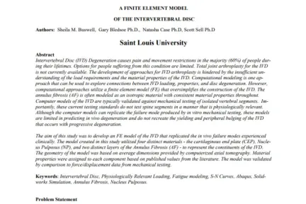
The aim of this study was to develop an FE model of the IVD that replicated the in vivo failure modes experienced clinically. The model created in this study utilized four distinct materials - the cartilaginous end plate (CEP), Nucleus Pulposus (NP), and two distinct layers of the Annulus Fibrosis (AF) - to represent the constituents of the IVD. The geometry of the model was based on average dimensions provided by computerized axial tomography. Material properties were assigned to each component based on published values from the literature. The model was validated by comparison to force/displacement data from mechanical testing.
Customer Papers